
Bone research leads to savings and better welfare
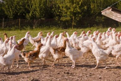
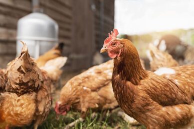
University of Alberta researchers are using a bone scanner to calculate the bone density in laying chickens, in the hope of finding ways to save the poultry industry money, while also providing better welfare for birds.
Dr Douglas Korver, a professor of poultry nutrition in the University of Alberta Department of Agricultural, Food, and Nutritional Science, hopes to find ways to prevent osteoporosis and bone breaks in laying hens.
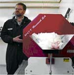
Korver and his team use a Quantitative Computed Tomography (QCT) to undertake their research.
“Laying hens put out an awful lot of calcium in the form of egg shells,” said Korver. That output of their bodies’ calcium can make their own bones as fragile as, well, egg shells, he said.
So far, Korver’s research points to a form of vitamin D that helps the chickens absorb more calcium, and demonstrating that allowing the birds to reach sexual maturity and start laying eggs later in life reduces the liklihood that bones will become fragile as the chickens age.
“If we can reduce the incidence of osteoporosis, for example, from 10 per cent of the flock to eight per cent of the flock, we’ve calculated that would save the Alberta industry about $160,000 a year, and the Alberta industry is only about 10 per cent of the Canadian industry.”
But the benefits do extend beyond financial considerations. In the past, the only way to take a look at bone development in chickens involved killing the birds. For a QCT scan, however, the hen is simply anaesthetised so it remains motionless for the 20-minute scan, then gradually regains consciousness.
“We are reducing the number of birds we have to put in an experiment at all, and we’re just getting a whole lot more data out of each bird,” said Korver.
Keeping the chickens alive for the scanning process also means having a constant data source and fewer variables when it comes to collecting information.
“The biggest thing that we’ve been able to do is follow the progression of a single bird throughout its life cycle,” said Korver. “We can relate feed intake to that particular point in its life and we can relate egg production to that particular bird.”
The scanner was provided to the university and is owned by DSM Nutritional Products.
Join 31,000+ subscribers
Subscribe to our newsletter to stay updated about all the need-to-know content in the poultry sector, three times a week.


 Beheer
 WP Admin
  Bewerk bericht