
Identification and speciation of oocysts in turkeys
Location of the lesions in the intestine helps in identification and speciation of Eimeria oocysts. Without any history of gross lesions, the speciation of oocysts cannot be determined solely based on the size of the oocysts, as there is an overlap in oocyst size and shape between different species. The availability of molecular diagnostic tools, such as PCR and sequencing, helps in discerning the Eimeria species.
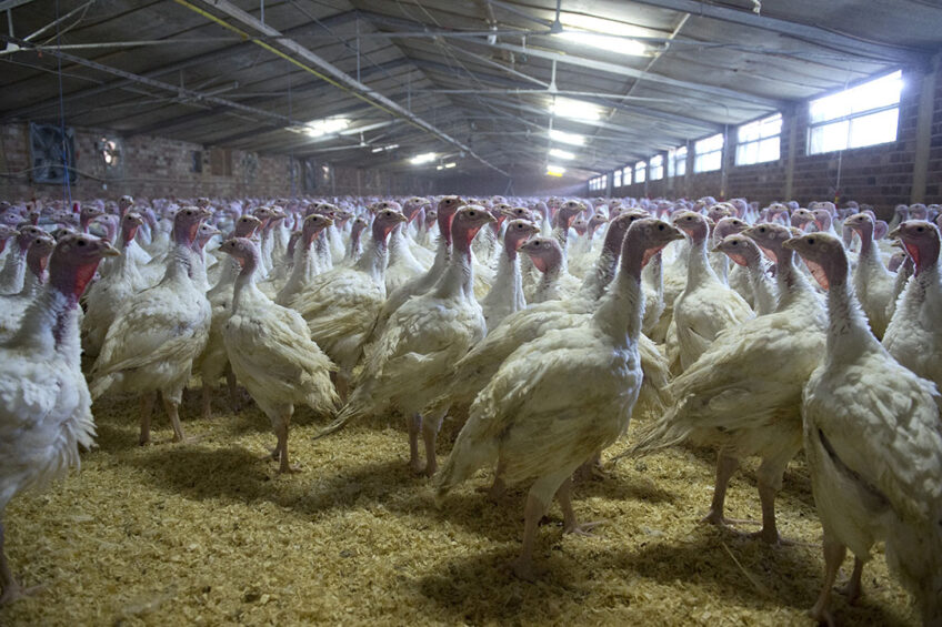
Coccidiosis is one of the most economically detrimental diseases in the turkey industry. Coccidiosis is caused by Eimeria species, a common intracellular protozoon. In commercial production systems, turkeys may be screened by faecal flotation or mucosal intestinal scrapings at regular intervals to identify Eimeria oocysts.
An increase in oocyst count during faecal flotation, especially if associated with clinical signs, raises concerns about turkey coccidiosis. In this diagnostic report, faecal samples from a 7-week-old turkey flock were investigated following an increase in the number of Eimeria oocysts detected. Faecal flotation followed by DNA extraction, PCR and sequencing, confirmed the oocysts as E. meleagrimitis, a highly pathogenic strain.
Ubiquitous
Intracellular protozoan parasites of the genus Eimeria are ubiquitous and prevalent in intensive turkey production facilities. They affect the production and performance of turkeys and significantly impact profitability. Economic losses are not just attributed to mortalities, but include poor feed conversion efficiency, lowered body weight, non-uniform sized birds, vaccinations, treatment and medications. Based on the 2022 turkey industry annual report, turkey coccidiosis ranks number 9 among the top health issues of the US turkey industry. A total of 7 Eimeria species, including E. adenoeides, E. gallopavonis, E. meleagrimitis, E. dispersa, E. meleagridis, E. innocua, and E. subrotunda, are found in turkeys.
Turkey coccidiosis is controlled and managed with anticoccidial programmes that include ionophores, in-feed medication, vaccination or phytonutrient supplements. In commercial turkey production systems, samples collected from the flock may be screened at various points in time.
An increase in the number of oocysts observed raises concerns about coccidiosis. In such cases, it may be important to know which Eimeria species is involved. In general, gross lesions of turkey coccidiosis are not as pronounced as in chickens. In cases of clinical coccidiosis, the gross lesions serve as a guide to determine the Eimeria species based on pathological manifestations. However, subclinical coccidiosis does not induce classical lesions of specific Eimeria species in the targeted organs but still affects production parameters.
Identification and speciation of turkey Eimeria species can be done based on the location of lesions in the intestine, along with the size and shape of the oocysts. In instances where gross lesions are not evident and with mixed infections, identification of Eimeria species oocysts is primarily done based on the size and shape of the oocysts with advanced molecular tools like PCR and sequencing.
Case report
In December 2022, faecal samples were submitted from a 7-week-old-turkey flock with a history of enteritis and a slight increase in mortality. Coccidiosis was suspected based on the detection of an increased number of Eimeria oocysts at different points in time with an in-house flotation test. Faecal samples were collected and submitted to investigate and determine the species of the Eimeria oocysts.
Screening of faecal samples with faecal flotation
Fecal samples were diluted in saturated sucrose solution and strained using 100 µm cell strainer (Corning). The filtrate (500 ul) was placed on McMaster slides and allowed to sit for 30 minutes and then examined under the microscope. DNA was extracted by PCR from 200uL of post-faecal float material using QIAamp Fast DNA Stool Kit following the manufacturer’s guidelines with the addition of using glass beads to rupture oocysts, along with 30 µl of proteinase K (Qiagen).
Each 25 µL PCR reaction consisted of 1X GoTaq G2 Hot Start Master Mix, 0.4 µM of each primer, and 5 µL of template. PCR was performed against the mitochondrial cytochrome c oxidase subunit I (mtCOI) gene of turkey Eimeria species based on the reference publications. On microscopic investigation of faecal flotation samples in the McMaster chamber, Eimeria oocysts were detected. In the PCR performed 6 Eimeria species (E. adenoeides, E. gallopavonis, E. meleagrimitis, E. dispersa, E. meleagridis, and E. innocua) amplicons were generated only with the primers targeted on E. meleagrimitis and confirmed by sequencing.
Finding the culprit
Eimeria species are strictly host and site specific. Location of the lesions in the intestine helps with the identification and speciation of Eimeria oocysts. However, in this study, faecal samples were investigated without any history of gross lesions. Even though faecal flotation revealed Eimeria oocysts in the faeces, the species of oocysts cannot be determined solely based on the size of the oocysts as there is an overlap in oocyst size and shape between different species (Table 1). The availability of molecular diagnostic tools, such as PCR and sequencing, helped in determining the Eimeria species.
Among Eimeria species, 3 are considered highly pathogenic, including E. meleagrimitis, E. adenoeides and E. gallopavonis. E. meleagrimitis affects the duodenum and causes watery and mucoid content, fibrin strands, small blood clots, hemorrhage, diphtheritic membranes and necrosis. With severe infection, the above lesions extend to the jejunum. E. meleagrimitis infections cause clinical signs such as listlessness, dehydration, diarrhea with mucus or blood-tinged faeces.
It should be noted that the presence of few oocysts in the faecal sample does not confirm that the flock has clinical coccidiosis. Clinical coccidiosis is accompanied by increased mortalities, classical lesions in the target organs and decreased production performance. Subclinical coccidiosis does not induce high mortalities or classical lesions in the target organs, but still affects production performance.
Although the location of the lesions in the intestinal tract was unknown, faecal flotation along with PCR and sequencing helped with the identification and speciation of E. meleagrimitis oocysts in this turkey flock.
Join 31,000+ subscribers
Subscribe to our newsletter to stay updated about all the need-to-know content in the poultry sector, three times a week.


 Beheer
 WP Admin
  Bewerk bericht