
Understanding chicken vision without the use of MRI
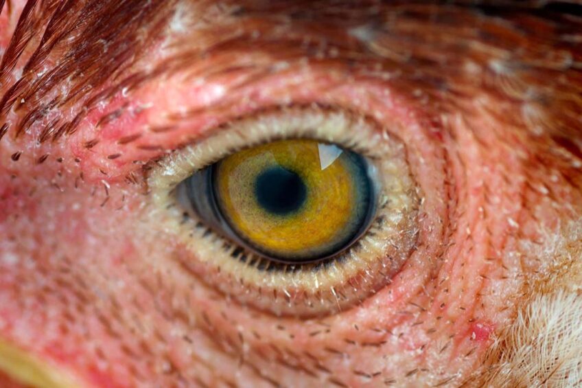
US scientists have developed 3D anatomy tech to learn more about chicken vision without having to use more expensive magnetic resource imaging (MRI).
Poultry researchers at the Arkansas Agricultural Experiment Station mapped the intricate neurological pathways that control vision in chickens with detailed 3D models of the connections between the eyes and 4 regions of the brain.
Wayne Kuenzel, professor of physiology and neuroendocrinology at the Department of Poultry Science, said as well as being cheaper than using MRI technology, the method would benefit teaching complex anatomy and expand the tools of animal science researchers.
“What is important about this technique is that it is a straightforward procedure, and it is not expensive. I am sure it will gain importance over time and attract a greater audience. The tectofugal visual pathways has 4 critical neutral structures in 4 different brain regions. Diagramming them in 3D enables one to see the entire pathway in 1 image and therefore should enable the learning of the entire pathway more rapidly and perhaps more permanently,” he said.
Using iodine to stain tissue
The scientists created the new 3D imaging by combining a conventional imaging method called histochemistry with a newer image method known as diceCT, which stands for diffusible iodine-based contrast-enhance computer tomography. Histochemistry uses chemical reagents like dyes to stain tissue and allow it to undergo image analysis. DiceCT is like an MRI, but instead of using a large magnet and radio waves, it uses iodine to stain the tissue so that the viewer can see groups of cells among fibre tracts. DiceCT uses x-ray scans to “digitally” slice the biological subject being studied.
Principal author of the study, Parker Straight, said the hybrid method of 3D scanning could be used to study neurobiology at a large scale, such as brain region morphology, and at a more detailed scale, such as looking at a single neurological pathway. An example of the technology’s potential use would include assessing changes or lesion patterns at various stages of disease.
Other examples, said Straight, might include long-distance neuron tracing without cutting the connection, as well as comparing structural differences and how they relate to different behavioural patterns. “The list is quite long in terms of how this method can be proven beneficial to research. I hope this study will prompt more investigations of animal neurobiology using 3D methods and how it compares to neurobiology of humans.”
If researchers wanted to implement the exact imaging pipeline the bird would have to be euthanised, but the diceCT portion of the imaging method could be done in live animals if they are sufficiently sedated so that a researcher could capture a clean 3D scan.
*The paper ‘Mapping the avian visual tectofugal pathway using 3D reconstruction’ has been published in the Journal of Comparative Neurology.
Join 31,000+ subscribers
Subscribe to our newsletter to stay updated about all the need-to-know content in the poultry sector, three times a week.


 Beheer
 WP Admin
  Bewerk bericht